Where Does an Image Become a Photograph? — When Microscopy Enters a Photography Museum
Update (October 12, 2026): On October 9, 2026, Nikon disqualified the first-place Small World in Motion 2026 video for not complying with its rules on generative AI, and said it would revisit its rules and evaluation procedures. Why Was a Winning Microscopy Video Disqualified? looks at this, including the absence of any clause in the current Nikon Small World rules asking entrants to disclose processing. The text below remains as of July 28, 2026.
A cell membrane glows blue against a dark field. Fine blood vessels branch like winter trees; a tissue section begins to resemble an unfamiliar landscape. In a laboratory, those colors and forms have names and an observational purpose. In a photography museum, they first meet us as images.
On July 25, 2026, the Tokyo Photographic Art Museum opened Nikon Small World / NIKON JOICO AWARD: The Microscopic World — Illuminating the Future of Life. Alongside recent prize-winning images, the exhibition includes historical microscopes, design records, and a daguerreotype microscopy image. It follows the development of the microscope and the images it produced from both scientific and artistic directions.
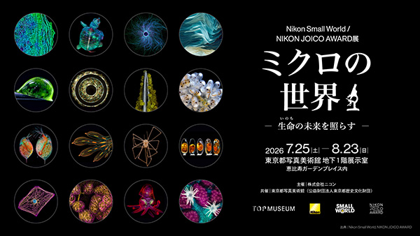
When does a microscopy image become a photograph? When the exposure is made? When the file is processed? When it wins an award? Or when it is placed on the wall of a photography museum?
The short answer
A microscopy image does not become a photograph only after it enters a museum. The moment someone chooses where to look within a specimen, selects the light, sets focus, and records the image, it already contains decisions made by its maker.
What the museum changes is not so much the image as the viewer's role. In the laboratory, the question is, "What can we learn from this?" In the gallery, we can also ask, "Why is it difficult to look away?"
That does not mean the line between science and art should be dissolved. The presence or absence of processing is less useful than whether we can trace what was made visible and how. A microscopy image may be scientific evidence and a photograph to be viewed. Process records and clear explanation allow it to carry both roles.
Scope of this article
The author has not yet seen the exhibition in full. This is a pre-visit note based on the Tokyo Photographic Art Museum's exhibition page, Nikon's public materials, and competition rules available on July 28, 2026.
The wall labels and research summaries must still be checked in person to understand the specimen, imaging method, and processing behind each work. This is not a review of individual images. It is a framework for looking at microscopy as photography.
The same image does different work in a laboratory and a museum
The exhibition has three sections. The first traces the history of microscopy. The second presents Nikon Small World and NIKON JOICO AWARD winners. The third looks at current uses of microscopes in science, medicine, and industry. The winning images and films in the second section are arranged along the evolution of life, from algae and plants to animal cells.
The structure matters because it does not isolate the winners as merely strange and beautiful views of a miniature world. The history of the instruments comes before them, and their present-day applications come after. Viewers can move between how the image looks, how it was made, and what it has been used to understand.
In a laboratory, microscopy images support observation, recording, measurement, and comparison. In a gallery, the same image can also be read through repeated forms, layered color, and depth. Its scientific meaning has not vanished. The pressure to reduce it immediately to one meaning has.
The wall does not turn the image into art by decree. It gives the image another kind of time.
Microscopy also has an outside to the frame
It is easy to imagine a microscopy photograph as a machine-made copy of truth. Yet it contains many choices familiar from other forms of photography.
Which specimen should be prepared? Which area deserves greater magnification? What illumination and imaging method should be used? Where should focus fall, and when should the image be recorded? If several focal planes or fluorescence channels are combined, which information belongs in the final frame? What is not selected remains outside the microscope's field of view.
The JOICO AWARD application asks for the title and paper information as well as details of the sample, microscope, and observation method. Nikon Small World's official rules require an image made by the entrant through a light microscope using a microscope objective, together with equipment and image descriptions.
The microscope makes the invisible visible, but the resulting image is not a neutral window. It has passed through specimen preparation, an optical system, and a series of human decisions. In that sense, microscopy has composition, exposure, and a world outside the frame.
Does processing make an image unscientific?
Processing alone does not tell us whether a scientific image is accurate.
The 2026 NIKON JOICO AWARD entry guide accepts microscopy images processed or altered with software. It also permits several source images to be assembled as a single montage. When processing has been used, however, the entrant must describe what was done in the work information.
Some microscopy techniques require signals beyond ordinary vision to be translated into a form that can be distinguished. Brightness and contrast may be adjusted, colors may be assigned to separate fluorescence signals, and several focal planes may be integrated. Processing can be part of making an observation legible.
The problem begins when processing changes the meaning of the data. AMED's research-integrity guide to appropriate image processing explains that suitability depends on the research purpose and analytical method. It also stresses retaining original images and related data, documenting software and procedures, and applying equivalent adjustments across the whole image and comparable controls. Removing only inconvenient details or treating comparison images differently is more than a matter of appearance.
Darkroom and software adjustments are common in art photography too. The distinction is not simply whether someone touched the image. In science, it must remain possible to test whether that touch distorted the observation. Beauty and accuracy can coexist, but accuracy needs a record.
One image is not enough to become a “research-result work”
The most revealing feature of the JOICO AWARD is that an entrant cannot submit a microscopy image by itself.
Three items are submitted together as one research-result work:
- a microscopy image;
- a visual research overview written for a general audience;
- a published academic paper explaining the image's scientific background.
The microscopy image and overview are judged for both artistic and academic merit; the paper is judged for academic merit. Artistic and academic scores carry equal weight. The published criteria for art include creativity, beauty, impact, technical command, and clarity. Academic criteria include novelty, originality, importance, universality, and contribution.
Science is not merely decorating itself with art here, nor is an artwork borrowing scientific language for authority. The image catches the eye, the overview opens a path into the research, and the paper shows where that image stands within a body of knowledge. Each has a different task.
A published paper does not automatically prove that every operation performed on the submitted image was correct. It establishes the research background. Assessing the image itself may still require the original data, acquisition settings, and processing record. The three-part structure is strong, but it is not a substitute for verification.
This distinction also helps in the museum. If we stop at "beautiful," the research subject becomes a pattern. If we read only the explanation, the reason to spend time with the image disappears. The context is there to let the viewer move back and forth.
Is there one author of a scientific photograph?
In ordinary photography, the person who released the shutter is usually named as the author. In microscopy, that outline is less tidy.
One person may prepare the specimen, another design the stain or fluorescent label, another operate the microscope, and someone else set the capture parameters or process the result. The research itself may have been conceived by a wider team. The authors of the paper are not necessarily the same person who acquired the image.
The JOICO AWARD accepts individual and group entries, with one representative named for a group. At the same time, its rules require the entrant to hold ownership and copyright and to obtain prior consent when the work comes from research conducted with a third party. A competition needs a definite rights holder. That administrative need does not reduce the making of the image to one pair of hands.
When reading an author's name in the gallery, it is worth asking not only who pressed the capture button, but who made the decisions that allowed the image to exist. Information about collaborators, specimen preparation, and processing is part of how the work should be seen.
Owning while opening
The JOICO AWARD applies a CC BY 4.0 license to submitted microscopy images. Entrants retain copyright. Others may share and adapt the image, including commercially, if they provide attribution and the license, and identify changes.
That is not the same as giving the work away. Authorship and copyright remain. What changes is that some control over reuse is opened in advance. Under Creative Commons Attribution 4.0, people who follow the terms may copy, redistribute, remix, and adapt the material.
Research can be opened while a work remains owned. Ownership here is not only a fence that keeps others out; it becomes the point from which the work can circulate while retaining the name of the person or team responsible for it.
There is still some tension. Science values openness for reuse and verification. Art markets may attach value to scarcity and controlled reproduction. JOICO images move between those systems. Even on a museum wall, they do not remain confined to that wall.
What to look for in the gallery
Alongside the beauty of the images, these are the questions I would bring to the exhibition:
- Are the colors observed colors, or were they assigned to distinguish signals?
- Is the work one exposure, or an integration of several focal planes or source images?
- How much is explained about the specimen, imaging method, processing, and research purpose?
- Is the credited author an individual or a team, and can the roles of collaborators be understood?
- How close are the research overview and paper to the displayed image?
- When historical microscopy images and recent winners are placed together, how do the standards of accuracy and beauty appear to change?
These questions do not dispel the image's magic. Knowing the marks left by its making keeps us from consuming its beauty as a decorative accident of nature.
Photography begins in more than one place
A microscopy image made in a laboratory may first serve as observational record or data. In a competition, it can become a research-result work accompanied by explanation and a paper. In a museum, it becomes a photograph that viewers can meet at their own pace.
This is not a one-way transformation. Scientific responsibility remains in the museum, and the author's name remains when a CC BY image circulates outside it. A single image can be evidence, explanation, and work without settling into only one of those roles.
Where, then, does it become a photograph? Probably not at the instant an institution declares it one. Photographic work begins when a portion of the world is separated by light, translated into a visible form, and left with enough information for others to trace that translation.
The museum does not decide where that beginning lies. It returns time for looking to an image first made elsewhere.
Related articles
Sources and verification
- Tokyo Photographic Art Museum: Nikon Small World / NIKON JOICO AWARD exhibition — dates, curatorial premise, three-part structure, historical microscopes, and the daguerreotype microscopy image.
- Nikon Healthcare Business Unit: exhibition announcement — exhibition structure, winning works, and the workflow from sample preparation to observation, capture, and analysis.
- Tokyo Art Beat: Nikon Small World / Nikon Joico Award Exhibition — secondary confirmation of dates and venue.
- NIKON JOICO AWARD: 2026 entry guide — the three-part submission, judging, processing disclosure, group entries, copyright, and CC BY.
- Nikon Small World: Photomicrography Competition Official Rules — light microscopy, microscope objectives, entrant-made images, and equipment descriptions.
- AMED: Appropriate Image Processing — scientific processing, original data, and processing records.
- Creative Commons: Attribution 4.0 International — sharing, adaptation, attribution, and change notices under CC BY 4.0.